
Rocabado TMJ Protocol
It includes a set series of TMJ Cone Beam takes in different joint positions. Provides the clinician with a complete joint view of the dynamics of both condyles.
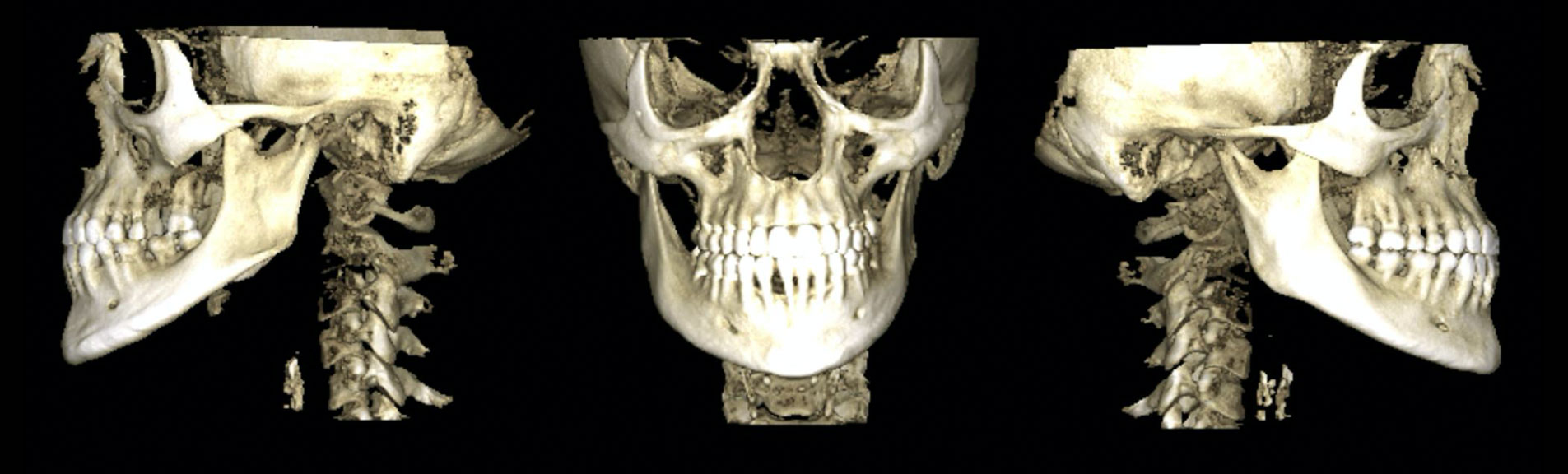
Craniovertebral Imaging
Craniovertebral Imaging
Use of 3D and 2D images to evaluate the stability of the cervical spine and the position of the skull in static and dynamic positions. Craniovertebral Study Dynamic Teleradiographies and Cone Beam Atlas-Axis
Cranium Teleradiographies
Cranium Teleradiographies
It is an X-ray of the skull and face taken at a distance. It is requested as a complementary examination in an orthodontic study and orthognathic surgeries.